37th ESP Annual Convention

Values-Driven Endodontic Practice
37th ESP Annual Convention | Values-Driven Endodontic Practice
Marco Polo Ortigas Manila, Pasig City

Speakers and Lectures

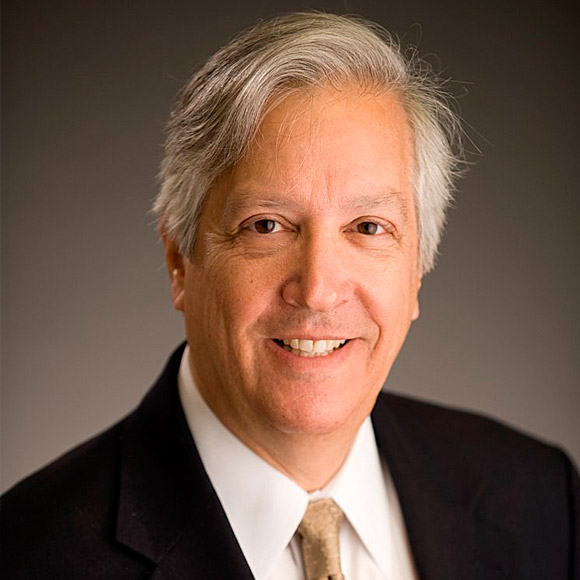
Arnaldo Castellucci, MD, DDS

Arnaldo Castellucci, MD, DDS

Dr. Arnaldo Castellucci graduated in Medicine at the University of Florence in 1973 and he specialized in Dentistry at the same University in 1977. From 1978 to 1980 he attended the Continuing Education Courses on Endodontics at Boston University School of Graduate Dentistry and in 1980 he spent four months in the Endodontic Department of Prof. Herbert Schilder. Since then, he has a limited practice to Endodontics.

He is an Active Member of the Italian Endodontic Society (SIE) where he worked as Scientific Advisor, Secretary Treasurer, Vice President and as President in 1993-1995. Dr. Castellucci is an Active Member of the European Society of Endodontology (ESE), he was the Secretary in 1981-1983. He has been the President of the International Federation of Endodontic Associations (IFEA) in 1990-1992. He is an Active Member of the American Association of Endodontists (AAE) since 1985.

He is an Assistant Professor of Endodontics at the University of Cagliari Dental School and Assistant Professor of Micro-Surgical Endodontics at the Specialty of Oral Surgery at the University of Naples Federico II.

He is the Editor of “The Italian Endodontic Journal” and of "The Endodontic Informer" and is Editor-in-Chief of Endo Tribune.

As an international lecturer, Dr. Castellucci gives presentations at National and International Congresses in 56 different countries. He has published more than 60 articles on Endodontics in the most prestigious Endodontic Journals and is the author of the text "EndodonticS", which is now available in three volumes in the english language. He is also the author of the textbook on Micro-Surgical Endodontics which will be published in the near future.

Dr. Arnaldo Castellucci is the Founder and President of the "Warm Gutta-Percha Study Club" and of the "Micro-Endodontics Training Center" in Florence, Italy where he teaches and gives hands-on courses.

Pre-Congress: Significance of Cavity Access and Contemporary Approach to Root Canal Shaping

Congress: Many Good News in Non-Surgical Endodontics!

Today, to perform a correct root canal treatment is easier and easier like never before. The powerful ultrasonic units and the ultrasonic tips enable also the general practitioner to prepare quickly and safely a correct access cavity. The finishing of the access cavity and the flaring of the walls and its cleaning are definitely steps that are facilitated by the use of this instruments. The identification of the 2° canal of the mesiobuccal root of the upper molars can be done without the risk of causing a perforation, eliminating in complete safety the portion of the mesial wall that hides its orifice. The calcifications of the pulpal floor that impairs visibility and therefore the identification of the openings and access to the root canals, can be easily removed with the ultrasonic tips, without changing the original anatomy and causing iatrogenic damages. Posts in carbon or glass fibre can be easily removed from within the canal using the self-cleansing ultrasonic tips. In conclusion with the ultrasonic tips the removal of metal posts both cast and prefabricated, is facilitated.

New exciting instruments became recently available to facilitate root canal preparation. Today it is fast, safe and easy to obtain the glide path using the new rotary NiTi files. They pre-enlarge the foramen to safely accommodate any NiTi System, avoiding the risk of straightening the curvatures, blocking the original canal, making ledges or fracturing the rotary file.

After this preliminary enlargement and obtaining the glide path, today the shape of the root canal can be obtained with the last generation of NiTi rotary files, to quickly prepare the canal to a three-dimensional obturation!

To this, must be added the results that can now be obtained thanks to the use of the operating microscope, revolutionary materials like Bioceramic sealers and the CBCT that gives the possibility to examine any tooth in three dimensions. All these innovations have the wonderful capability of making endodontic diagnosis more accurate, and endodontic treatments more predictable, easier, enjoyable and with a higher percentage of long term success.

Learning Objectives:

  • To make a more accurate diagnosis and a more accurate treatment plan
  • To make a precise access cavity and an easy identification of the canal orifices
  • To safely and quickly shape the root canal in the total respect of the original anatomy
  • To safely and quickly obturate the root canal system in the three dimensions
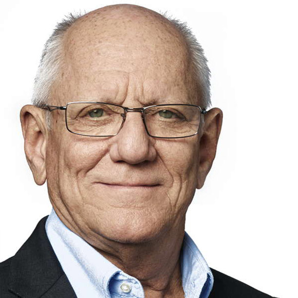
Alan Gluskin, DDS

Alan Gluskin, DDS

Dr. Alan Gluskin is currently Professor and Co-Chairperson, Department of Endodontics, Arthur A. Dugoni School of Dentistry, University of the Pacific in San Francisco, California. He is at present a Fellow of the International College of Dentists and the American College of Dentists.

Dr. Gluskin is a member of numerous societies including the American Association of Endodontists (AAE), the American Dental Association (ADA) and the San Francisco Dental Society. He has served on the Educational Affairs Committee and the Research and Scientific Affairs Committee of the AAE, as well as General Chair for AAE10 and AAE18.

Dr. Gluskin is presently on the Executive Board of Directors of the American Association of Endodontists as President-Elect of the AAE, and additionally functions on the editorial boards of the Journal of Endodontics and the International Journal of Endodontics. Dr. Gluskin is the 2017 recipient of the University of the Pacific’s Eberhardt Teacher-Scholar Award. He is co-editor of the textbooks Decision Making in Dental Treatment Planning and Practical Lessons in Endodontic Treatment.

Endodontics and Neurologic Injury

Neurologic injury as an outcome of endodontic treatment can have devastating consequences for both the patient and the endodontist. Mishaps that occur during the therapeutic stages of endodontic therapy can result in neurologic damage with an unpredictable course and an uncertain prognosis. Dr. Gluskin will discuss the evidence and best practices regarding neurologic impairment as a result of endodontic care. He will review the data for prognosis and how to determine treatment and intervention strategies in local anesthetic injury; heat and chemical harm from overfill; as well as hypochlorite accidents and the resulting neural trauma caused by surgical error.

Learning Objectives:

  • To recognize the advantages of new 3-D imaging technologies in identifying key neurovascular structures and their proximity to the root canal systems of teeth
  • To appreciate the hydraulic outcomes of common obturation methods and their potential for injury and mishaps
  • To identify safe protocols for avoiding extraradicular overfill of  the root canal space when there is close proximity to neurovascular anatomy
  • To be familiar with prognosis and best practice for injuries and mishaps that result in neurologic damage

Can Endodontically Treated Teeth Last a Lifetime?

New Frameworks for Advancing Structural Conservation in Endodontics

The primary goal of endodontic therapy is the long-term retention of a functional tooth by preventing or treating apical periodontitis. However, there are many other factors that impact endodontic outcomes such as the quality of the restoration and structural integrity of the tooth after root canal preparation. Contemporary research efforts are currently directed to better understanding dentin behavior and structure during aging and function. An alternative approach is to minimize structural changes during root canal therapy, which may result in a new strategy that can be labelled “minimally invasive endodontics”.

Learning Objectives:

  • To understand the structural weakening that occurs as a result of endodontic and restorative procedures
  • To reflect upon the principles of cervical dentin preservation in stabilizing load transfer to roots
  • To appreciate the biocompatibility of restorative materials and clinical strategies for protecting endodontically treated teeth
  • To learn how imaging technologies can inform the practitioner about structural liability
  • To recognize fracture susceptibility in all teeth and the predisposition for damage
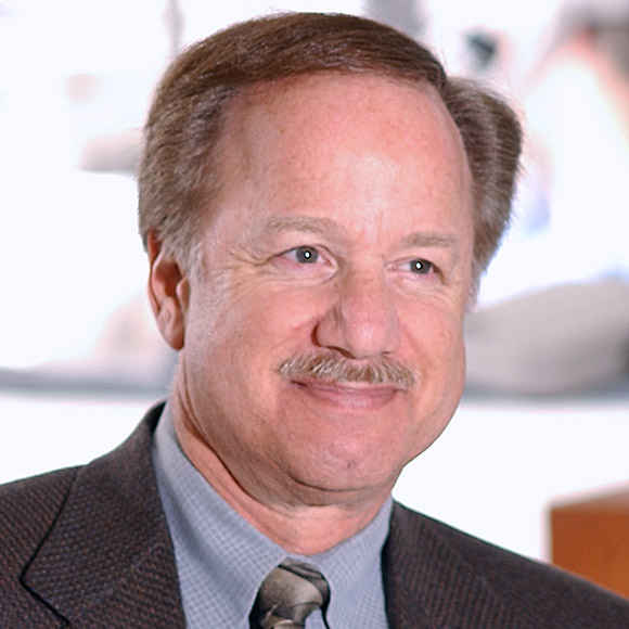
Martin Levin, DMD

Martin Levin, DMD

Dr. Martin Levin is Adjunct Professor of Endodontics and Chair of the Dean’s Council at the University of Pennsylvania. He has served as Co-Chair of the Joint Special Committee on the Use of Cone Beam Computed Tomography, American Association of Endodontists (AAE) and the American Academy of Oral and Maxillofacial Radiology. Dr. Levin is currently appointed to the AAE’s Special Committee to Develop an Outcomes Consensus Conference. He is a contributor to the Pathways of the Pulp, and Ingle’s Endodontics, as well as the author of numerous book chapters and peer-reviewed articles. Dr. Levin earned his degree in dental medicine and completed his endodontic training at the University of Pennsylvania. He is a Diplomate of the American Board of Endodontics.

Use of CBCT in Endodontics: What Every Dentist Should Know

Endodontic lesion and implant site assessments are important tasks in today’s dental office. Both tasks require diagnostic radiographs and image-guided treatment. Periapical and panoramic radiography have been augmented by the introduction of limited field of view (FOV) high-resolution CBCT, allowing three-dimensional assessment of odontogenic and non-odontogenic lesions, root canal morphology, revision treatment, root and alveolar fractures, resorptive lesions, anatomy prior to surgery and implant sites. This lecture will highlight the advantages of CBCT imaging for mapping of important anatomic structures and periapical lesion visualization.

Learning Objectives:

  • To list the benefits of limited FOV, high-resolution CBCT in identification of complex dental morphology
  • To describe how volumetric analysis can aid in the diagnosis of periapical pathosis when contradictory clinical signs and symptoms are present
  • To understand how volumetric analysis can improve the accuracy of perioperative measurements in the assessment of osteolytic lesions
  • To learn about the most current research supporting the use of CBCT in endodontic assessments
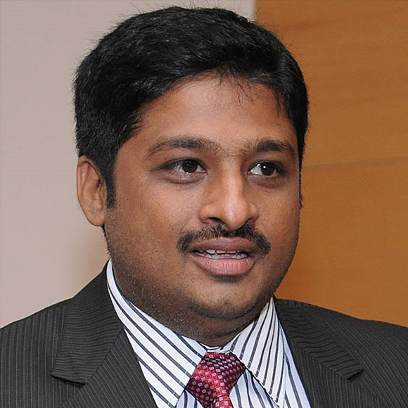
S Sai Kalyan, BDS, MDS

S Sai Kalyan, BDS, MDS

Dr. S Sai Kalyan is an academician with over 15 years of work experience (Associate Professor of Conservative Dentistry and Endodontics, RDC Pravara Institute of Medical Sciences, Lecturer at Vishnu Dental College, Andhra Pradesh, 2002-2003). He has closely worked with several medical device manufacturing companies as their Key Opinion Leader (Prevest Denpro Ltd. and Sybron Endo, USA). He is actively involved in quality control programs at the University and a member of the University IQAC team. He occupied various positions on the Board of studies and Academic Council, Pravara Institute of Medical Sciences. He is an active member of the University Ethical Team for Clinical trials and was the Secretary of the Institutional Ethical Committee. He is a member of the Indian Dental Association, World Association of Growth Factor and Stem Cells in Dentistry, Societa Italiana di Odontostomatologia e Chirurgia Maxillo Facciale, Italy; an International member of GEACoP, Brazil and Life member of Indian Association of Conservative Dentistry and Endodontics.

He received his MDS in Conservative Dentistry and Endodontics, RDC Pravara Institute of Medical Sciences Masters in Advanced Oral Surgery and Implantology from the University of Bari, Italy and BDS, from SDM College of Dental Sciences. He registered for PhD in Endodontics at Pravara Institute of Medical Sciences (his paper: "The Impact of Intracanal Nanoparticles and Photoactivated Disinfection on Endodontic Biofilm Bacteria").

He is a Fellow of the International College of Dentists, USA and Indian Board of Micro-Restorative and Endodontics; a Reviewer of Journal of Dental & Allied Sciences and Clinical Oral Insvestigations Editorial Board member of The Journal Of Conservative Dentistry, Journal of Dental & Oral Research, Journal of Oral Health and Dentistry, International Journal of Innovative Studies in Medical Sciences, International Journal of Growth factors and Stem Cells.

He has several articles published in peer-reviewed journals and has conducted several workshops.

Endovista: Netflix to Newer Technologies

Effectiveness of Root canal treatments is well established with a huge amount of published scientific data detailing its success. Lately, various holistic healers have created a non-scientific propaganda on the evil effects of endodontic treatment. Netflix aired a documentary titled “Root Cause” based on those non-scientific claims. The whole dental fraternity stood up for the specialty by sharing the scientific truth. The campaign led to the removal of the misinformation vehicle “Root Cause” from Netflix.

Endodontics has evolved tremendously and its applications have immensely improved the quality of dental treatment. The addition of regenerative endodontics has further widened the scope of treating pulpal diseases with Hence exploring the different aspects of the principles of endodontics, tackling certain challenges will help to boost its development with the advantage of innovations in research and technology.

Technological advances continue to make diagnosis and treatment more effective. Software improvements for Cone-Beam Computed Tomography systems (CBCT) are giving better views of canal systems and lessening artifacts. Advances in irrigation protocol are showing great improvements in our ability to debride and disinfect canal spaces while newer obturation materials are showing promise for broadening and lengthening our success rates.

Advances in endodontics has fundamentally changed our practice and resulted in reliable, efficient, fast and predictable endodontic care. A 3-dimensional approach towards pulpal diseases using proper biologic principles and advanced  biomaterials have changed the way we practice dentistry. This lecture outlines the scientific truths supporting endodontic therapy and discusses the various strategies which lay the foundation for Predictable 3D Endodontics.

Learning Objectives:

  • Myths and facts relating to the safety of endodontic therapy
  • Technological advancements in endodontics
  • Recent trends and newer biomaterials
  • Overview of Pulpal regeneration and bioactive restoratives
  • Novel therapies and predictable endodontic success
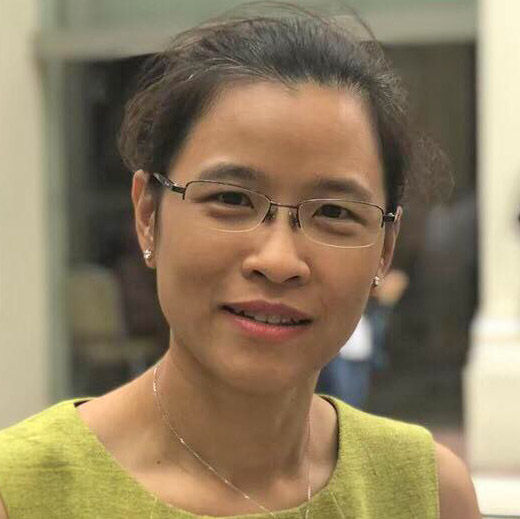
Somsinee Pimkhaokham, DDS, PhD

Somsinee Pimkhaokham, DDS, PhD

Dr. Somsinee Pimkhaokham is an Associate Professor at the Chulalongkorn University where she also finished her Doctor of Dental Surgery Degree. She acquired her Ph. D in Dental Science at the Tokyo Medical and Dental University, Japan under the Japanese Government Mambusho Scholarship for Post Graduates.

Dr. Pimkhaokham is a lifetime member of the Dental Association in Thailand since 1993 and the Thai Endodontic Association since 2000 and an Associate Member of the American Association of Endodontists.

Dr. Pimkhaokham has published in various journals locally and internationally.

Integrating Technology to Improve Treatment Outcome